Cartilaginous tissue: functions and features of the structure
It's no secret that athletes even in goodphysical form and at a relatively early age often give up training because of injuries. A large proportion of their problems are ligaments. The weakest part is cartilaginous tissue. The functions of damaged joints, it turns out, can be restored if timely attention is paid to the problem and to create suitable conditions for the treatment and regeneration of their cells.

Fabrics in the human body
The human body is a complex and flexiblea system capable of self-regulation. It consists of cells different in structure and function. In them, the main metabolism takes place. Together with non-cellular structures they are combined into tissues: epithelial, muscular, nervous, connective.
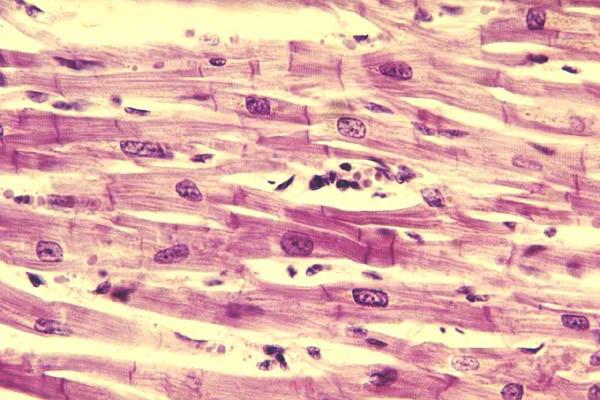
Epithelial cells form the basis of cutaneouscover. They lining the internal cavity (abdominal, thoracic, upper respiratory tract, intestinal tract). Muscular tissue enables a person to move. It also ensures the movement of internal environments in all organs and systems. The musculature is divided into the following types: smooth (walls of the cavity organs and vessels), cardiac, skeletal (transversely striated). Nerve tissue provides the transfer of impulses from the brain. Some cells are able to grow and multiply, some of them are capable of regeneration.
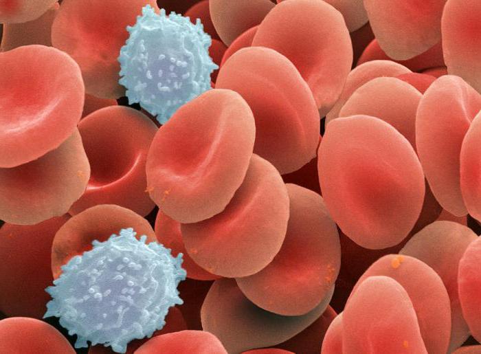
The connective tissue is the internal environmentorganism. It is different in structure, structure and properties. It consists of strong bones of the skeleton, subcutaneous fatty tissue, liquid media: blood and lymph. It also includes cartilaginous tissue. Its functions are formative, amortization, support and support. All of them play an important role and are necessary in a complex system of the body.
Cartilaginous tissue: structure and function
Its characteristic feature is looseness in locationcells. Considering them separately, you can see how clearly they are separated from each other. The intercellular substance - a matrix acts as a ligament between them. And in different types of cartilage it is formed besides the main amorphous substance by various fibers (elastic and collagen). Although they have a common protein origin, they differ in properties and, depending on this, they perform different functions.

All bones of the body were formed from cartilage. But as they grew, the intercellular substance was filled with crystals of salts (mainly calcium). As a result, the bones gained strength and became part of the skeleton. Cartilages also perform supporting functions. In the spine, being between segments, they perceive constant loads (static and dynamic). Ears, nose, trachea, bronchi - in these areas tissue plays a more formative role.
Growth and nutrition of the cartilage is carried out throughperichondrium. It is in the tissue is an obligatory part, except the joints. In them, between the rubbing surfaces there is a synovial fluid. It washes, lubricates and nourishes them, diverts the exchange products.
Structure
In the cartilage there are few cells capable of division, and manyspace around them, filled with various protein properties. Because of this peculiarity, the regeneration processes often take place in the matrix in a greater degree.
There are two types of tissue cells: gonrotsity (mature) and chondroblasts (young). They differ in size, place and method of location. Chondrocytes have a rounded shape, and they are larger. They are arranged in pairs or in groups of up to 10 cells. Chondroblasts are usually smaller and located in the tissue along the periphery or singly.
In the cytoplasm of cells under the membrane accumulateswater, there are inclusions of glycogen. Oxygen and nutrients enter the cells diffusely. There is a synthesis of collagen and elastin. They are necessary for the formation of intercellular substance. From its specifics, it depends on what type it will be cartilaginous tissue. Features of the structure and function of the larynx differ from the intervertebral discs, including the content of collagen. In the auricles, in the cartilage of the nose, the intercellular substance consists of 30% elastin.
Kinds
How is the cartilaginous tissue classified? Its functions depend on the predominance of specific fibers in the matrix. If there is more elastin in the intercellular substance, the cartilaginous tissue will be more plastic. It is almost as strong, but the fiber bundles are thinner in it. They well withstand loads not only on compression, but also on tension, capable of deformations without critical consequences. Such cartilage is called elastic. Their tissues form the larynx, ears, nose.
If the matrix around the cells is largecollagen with a complex structure of the construction of polypeptide chains, this cartilage is called hyaline. It often covers the inner surfaces of the joints. The greatest amount of collagen is concentrated in the surface zone. It plays the role of a skeleton. The fiber bundles in it resemble in structure the three-dimensional intertwined networks of a spiral shape.
There is one more group: fibrotic, or fibrous, cartilage. They, like hyaline, contain a large amount of collagen in the intercellular substance, but it has a special structure. The bundles of their fibers do not have a complex interlacing and are located along the axis of the greatest loads. They are thicker, have a special compressive strength, are poorly restored upon deformation. Intervertebral discs, joints of tendons with bones are formed from such tissue.

Functions
Thanks to the special biomechanical propertiesThe cartilage tissue is ideal for binding the components of the musculoskeletal system. It is able to accept the forces of compression and stretching during movements, redistribute them evenly to the load, to some extent absorb or dissipate.
Cartilages form abrasion-resistant surfaces. Together with the synovial fluid, such joints, under permissible loads, are able to perform their functions normally for a long time.
Tendons are not cartilaginous tissue. Their functions also consist in linking the musculoskeletal system into a common system. They also consist of bundles of collagen fibers, but their structure and origin are different. Cartilage of the nose, respiratory organs, and the auricles, in addition to performing the shaping and supporting functions, are the place of attachment of soft tissues. But unlike the tendons, the muscles next to them do not have this load.
Special Properties
There are very few vessels in the elastic cartilage. And this is understandable, because a strong dynamic load can damage them. How does the cartilaginous connective tissue feed? These functions take on the intercellular substance. In the hyaline cartilage there are no vessels at all. Their rubbing surfaces are quite tough and dense. They are fed by the synovial fluid of the joint.
In the matrix, water moves freely. It contains all the necessary substances for metabolic processes. Proteoglycanic components in the cartilage perfectly bind the water. It as an incompressible substance provides rigidity and additional depreciation. Under loads, water takes on an influence, spreads throughout the intercellular space and smoothly removes stress, preventing irreversible critical deformations.

Development
In the body of an adult up to 2% of the mass accounted foron cartilage tissue. Where is it focused and what functions it performs? Cartilage and bone tissue in the embryonic period is not differentiated. There are no bone embryos. They develop from cartilage and are formed at the time of birth. But part of it never ossifies. It forms the ears, nose, larynx, bronchi. It is also present in the joints of the arms and legs, joints of the pubic bones, intervertebral discs, meniscus knees.
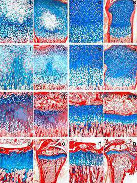
The development of cartilage occurs in several stages. At first, the cells of the mesenchyme are saturated with water, rounded, lose their processes and begin to produce substances for the matrix. After this, their differentiation into chondrocytes and chondroblasts occurs. The former appear to be tightly surrounded by intercellular substance. In this state, they can be divided a limited number of times. After such processes, an isogenic group is formed. Cells remaining on the surface of the tissue become chondroblasts. In the process of producing substances of the matrix, the final differentiation occurs, a structure is formed with a clear division into a thin border and the base fabric.

Age changes
Functions of human cartilage tissue in the process of lifedo not change. However, over time, signs of aging can be noticed: the muscles and tendons of the joints weaken, flexibility is lost, pain is disturbed due to changes in the weather or under unusual load. Such a process is considered the physiological norm. By the age of 30-40 years, the symptoms of changes may, to a greater or lesser extent, begin to cause inconvenience. Aging of the articular cartilage tissue is due to the loss of its elasticity. Loss of elasticity of the fibers. The fabric dries out, loosens.
Cracks appear on the smooth surfacebecomes rough. Smooth and easy sliding is no longer possible. The damaged edges grow, deposits are formed in them, osteophytes are formed in the tissue. Elastic cartilages age with the accumulation of calcium in the intercellular substance, but their functions (nose, auricles) are hardly reflected.

Impaired function of cartilage and bone
When and how can this happen? To a large extent, this depends on the function of the cartilage tissue. In intervertebral discs, the main function of which is stabilizing and supporting, most often the malfunction occurs during the development of dystrophic or degenerative processes. The situation may lead to displacements, which, in turn, will cause compression of the surrounding tissues. Inevitable swelling, pinching of the nerves, squeezing of blood vessels.
To restore stability, the bodytrying to deal with the problem. The vertebra at the deformation site “adjusts” to the situation, grows in the form of peculiar bone outgrowths (whiskers). It also does not benefit the surrounding tissues: again, swelling, pinching, compression. Such a problem is complex. Disorders of the bone and cartilage apparatus are called osteochondrosis.
Prolonged limitation of movement (gypsum withinjuries) also adversely affects cartilage. If, under excessive loads, elastic fibers are reborn into coarse fibrous bundles, then at low activity, cartilage ceases to eat normally. Synovial fluid does not mix well, chondrocytes receive less nutrients, and as a result, the required amount of collagen and elastin is not produced for the matrix.
The conclusion suggests itself: for normal functioning of the joints, cartilage should receive a sufficient strain on tension and compression. To ensure this, you need to exercise, lead a healthy and active lifestyle.